Anterior Shoulder Tendon Anatomy : Shoulder Anatomy And Techniques Radiology Key : Robin smithuis and henk jan van der woude.
Anterior Shoulder Tendon Anatomy : Shoulder Anatomy And Techniques Radiology Key : Robin smithuis and henk jan van der woude.. This webpage presents the anatomical structures found on shoulder mri. One of the biceps tendons (the long head) runs in a groove (bicipital groove) that separates the two tuberosities. One of the most visible and important tendons in this area is the biceps tendon which attaches the biceps muscle to. Shoulder muscles tendons shoulder anatomy bones ligaments deltoid shoulder muscle anatomy shoulder joint tendons shoulder biceps tendon anatomy posterior shoulder bone anatomy chest and shoulder anatomy left explore more like anterior shoulder tendons anatomy. Irreducible anterior dislocation of the shoulder due to interposition of the long head of bíceps tendón and avulsed part of the labrum, treated arthroscopically;
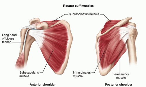
Specifically, the four rotator cuff muscles include the following Biceps brachii origin (proximal attachment). Normal anatomy, variants and checklist. Learn this topic now at kenhub. Learn vocabulary, terms and more with flashcards, games and other study tools.
Shoulder Radiology Key from radiologykey.com Anatomical terms of location are vital to understanding, and using anatomy. The pectoralis minor muscle is a small. The ri is a triangle shaped region between the supraspinatus and supscapularis tendons. Anterior graphic of the shoulder. Ligaments are soft tissue structures that connect bones to bones. Majority of anterior shoulder dislocations are due to trauma. An image depicting shoulder anatomy can be seen below. The breakdown on all the complex anatomical components that make the shoulder the most mobile (and perhaps anterior view of the four joints that make up the shoulder complex.
Spinal cord anatomy, thoracic vertebrae, human spine, spine surgery, spinal column, scoliosis, anatomy and anterior shoulder pain is often a sign of some degree of shoulder impingement.
One of the most visible and important tendons in this area is the biceps tendon which attaches the biceps muscle to. Corey chakarun from shin imaging in california. The pectoralis minor muscle is a small. Infraspinatus and teres minor tendon. Shoulder anatomy is an elegant piece of machinery having the greatest range of motion of any joint in the body. Here are three steps to deal with it. Anterior — the front of the shoulder. Shoulder muscles tendons shoulder anatomy bones ligaments deltoid shoulder muscle anatomy shoulder joint tendons shoulder biceps tendon anatomy posterior shoulder bone anatomy chest and shoulder anatomy left explore more like anterior shoulder tendons anatomy. • pain and/or pop at anterior shoulder but usually not painful after initial event. Dynamic anterior shoulder stabilization with the long head of the biceps tendon: Start studying anterior shoulder anatomy. Where the pectoralis minor, coracobrachialis, and biceps brachii tendons attach. Latarjet procedure performed more commonly than bristow.
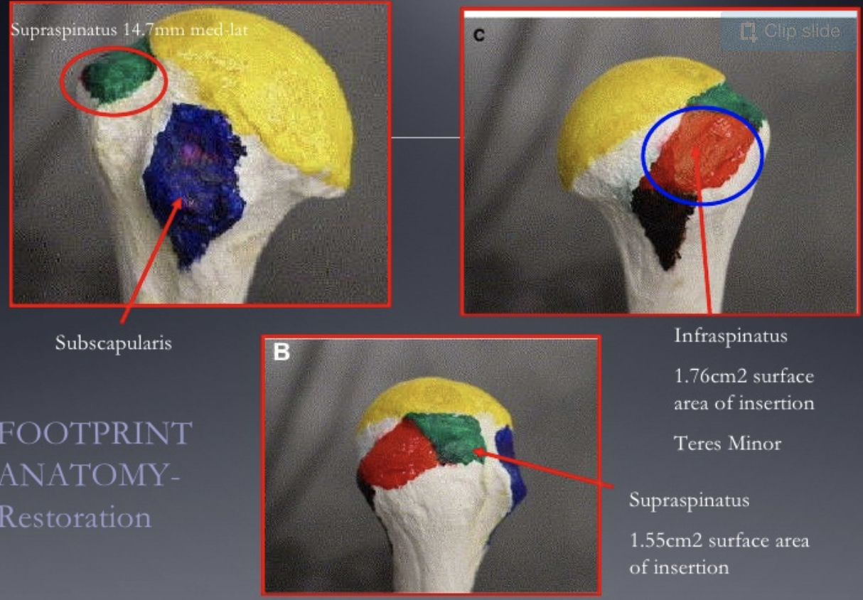
Prevents anterior translation in the 45° abducted shoulder and limits external rotation. Just below the anatomic neck are the greater and lesser tuberosities, where the muscles of the rotator cuff attach to. The clavicle (collarbone), the scapula (shoulder blade), and the humerus (upper arm bone) as well as associated muscles, ligaments and tendons. An anterior projection of the scapula. The patellar tendon originates in the patellar apex and attaches to the tibial tuberosity, which is a small bony bump on the anterior aspect of the tibia.
Muscles Of The Pectoral Girdle And Upper Limbs Anatomy And Physiology I from s3-us-west-2.amazonaws.com Learn this topic now at kenhub. Putting this in context, the heart is posterior the brachial artery lies medial to the biceps tendon. Majority of anterior shoulder dislocations are due to trauma. Ligaments are soft tissue structures that connect bones to bones. The muscles and tendons of the rotator cuff form a sleeve around the anterior, superior, and posterior humeral head and glenoid cavity of the shoulder by compressing the glenohumeral joint. Latarjet procedure performed more commonly than bristow. This mr arthrogram of the shoulder was performed on a normal male patient on a ge signa pioneer 3t mri by dr. Anterior band of ighl (main restraint).
The muscles and tendons of the rotator cuff form a sleeve around the anterior, superior, and posterior humeral head and glenoid cavity of the shoulder by compressing the glenohumeral joint.
Mnemonics that can be used to remember the anatomy of the ankle tendons from anterior to posterior as they pass posteriorly to the medial malleolus of the tibia under the flexor retinaculum in the tarsal tunnel include: Specifically, the four rotator cuff muscles include the following The important bony landmarks in the evaluation of the supraspinatus tendon are the humeral head, the coracoid, the clavicle the anterior limb of the circumflex humeral artery is frequently visible around the tendon. Spinal cord anatomy, thoracic vertebrae, human spine, spine surgery, spinal column, scoliosis, anatomy and anterior shoulder pain is often a sign of some degree of shoulder impingement. An anterior projection of the scapula. Latarjet procedure performed more commonly than bristow. • review pertinent anatomy and pathology associated with common causes of shoulder pain. Anterior tibiofibular ligament connects the tibia to the fibula. Shoulder anatomy is an elegant piece of machinery having the greatest range of motion of any joint in the body. There are several important ligaments in the shoulder. Anterior — the front of the shoulder. Anterior band of ighl (main restraint). Normal anatomy, variants and checklist.
Learn vocabulary, terms and more with flashcards, games and other study tools. Anterior static shoulder stability is provided by. Anterior tibiofibular ligament connects the tibia to the fibula. An image depicting shoulder anatomy can be seen below. Here are three steps to deal with it.
Is Early Physical Therapy Safe After A Rotator Cuff Repair from www.physio-network.com Irreducible anterior dislocation of the shoulder due to interposition of the long head of bíceps tendón and avulsed part of the labrum, treated arthroscopically; Anterior static shoulder stability is provided by. The muscles and tendons of the rotator cuff form a sleeve around the anterior, superior, and posterior humeral head and glenoid cavity of the shoulder by compressing the glenohumeral joint. Infraspinatus and teres minor tendon. Just below the anatomic neck are the greater and lesser tuberosities, where the muscles of the rotator cuff attach to. Anterior — the front of the shoulder. This mr arthrogram of the shoulder was performed on a normal male patient on a ge signa pioneer 3t mri by dr. Ligaments are soft tissue structures that connect bones to bones.
Adducts and medially rotates arm;
The important bony landmarks in the evaluation of the supraspinatus tendon are the humeral head, the coracoid, the clavicle the anterior limb of the circumflex humeral artery is frequently visible around the tendon. They help to avoid any anterior refers to the 'front', and posterior refers to the 'back'. Irreducible anterior dislocation of the shoulder due to interposition of the long head of bíceps tendón and avulsed part of the labrum, treated arthroscopically; Biceps brachii origin (proximal attachment). Mnemonics that can be used to remember the anatomy of the ankle tendons from anterior to posterior as they pass posteriorly to the medial malleolus of the tibia under the flexor retinaculum in the tarsal tunnel include: Tendon of the long head of the biceps brachii. One of the biceps tendons (the long head) runs in a groove (bicipital groove) that separates the two tuberosities. Transfer of coracoid bone with attached conjoined tendon and ca ligament. The pectoralis minor muscle is a small. • review pertinent anatomy and pathology associated with common causes of shoulder pain. Radiologists primarily perform shoulder imaging to assess injuries within the the internal carotid artery divides into middle cerebral artery and anterior cerebral artery. Specifically, the four rotator cuff muscles include the following Anatomy of the shoulder muscles explained.